
Description
Discover high-quality ultra-widefield retinal imaging
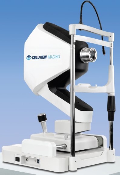
WRI-1: Introducing accessible high-definition ultra-widefield fundus imaging.
The WRI-1 Ultra-Widefield Retinal Imager allows for a comprehensive view of the retina, producing an up to 200° image via a two-image auto-stitch.
The steerable internal fixation facilitates image capture within the extended periphery encompassing regions beyond the traditional 200-degree field of view.
Comprehensive View of the Retina
It enables doctors to detect a wide range of retinal conditions and pathologies, including peripheral abnormalities that might otherwise go unnoticed with narrower field-of-view imaging.
By capturing an extensive retinal area, doctors can identify early signs of retinal diseases, such as diabetic retinopathy, retinal tears, or other peripheral retinal lesions, at a stage when intervention and treatment are most effective.
Patient Workflow and Efficiency
Non-mydriatic imager
The WRI-1 features a patient friendly low intensity flash system enabling imaging through pupils as small as 2.5mm. It improves patient flow by assisting operator activities, eliminating the need for dilating patients.
Multimodal imaging
The device has the ability of covering the full visible spectrum for Full Color and Infrared retinal imaging. Numerical filters are also available allowing different views of the retina.
Assisting in the detection and management of vision-threatening pathologies
Full-Color and Infrared Imaging
The full visible spectrum LED array offers full-color and infrared images for the detection of potential pathologies that may go unnoticed with other narrower imaging methods.
Nominal Staff Training
The WRI-1 is an easy-to-operate device even for unskilled operator usage. It presents an intuitive user interface and provides patient-friendly capture for a greater practice throughput.
Precision Imaging with Auto Focus & Auto Gain
Automatically adjusts focus and gain for sharp, high-resolution retinal images, streamlining the imaging process and reducing the need for manual adjustments by clinicians. The Auto Gain Control (AGC) is a closed-loop feedback that helps to maintain a constant image output irrespective of the patient’s pigment.
Unlimited Cloud Storage & Remote Access
Data storage and review
Unlimited cloud-based storage securely stores your valuable data in the cloud, accessible whenever and wherever you need it. Combining with the unlimited Remote Review stations it supports seamless data exchange for remote patient monitoring and collaborative eye care.
Remote service feature
Possible remote support when needed. Minimizing downtime and costs.
Automatic software and feature upgrades
Stay at the forefront of technology with seamless updates, ensuring you always have the latest tools readily available.
Technical Specifications
| Field of view | Single image: 133° / Two auto stitched images: 200° x 133° |
| Illumination source | 4 LED array covering 460-830nm |
| Image capture modes | Full color / Red-free / Infrared |
| Minimum pupil size | Non-mydriatic 2.5mm |
| Auto-focus | -15D to +15D (in steps of 0.25D) / Manual control available |
| Working distance | 12 mm |
| Pixel pitch resolution | 12 µm |
| Fixation target | Internal OLED, fixed points or user controlled across 133° FOV |
| Display | 22” high resolution LG monitor with FHD and/or 4K |
| Data storage | Unlimited patient data and images in the Cloud |
| Remote review stations | Unlimited review stations software included |
| System size (WxHxD) | 320 mm x 540 mm x 390 mm |
| Weight | 14 kg (30.9 lbs) |
| Electrical class | IEC60601-1 Class 1 |
| Power supply | 100-240 VAC / 24 VDC / 60 W Max |


